Advantages of the 3Shape X1
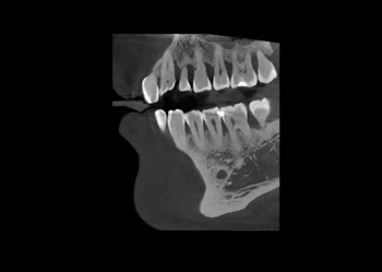
Pioneering technology enables uniquely sharp images
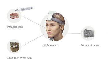
Digital 360-degree patient recordings

Offer your patients more services with an enhanced product
A CBCT scanner makes it possible to create a detailed, high-resolution image of the upper and lower jaw, temporomandibular joints and cavities as well as the dentition with 2 or 3D. With a CBCT scanner, dentists have the option of imaging teeth and surrounding bones in three dimensions, which gives the patient a much better diagnosis and a higher precision in treatment. 3D X-ray images can be shown directly to the patient, which leads to higher treatment acceptance and, in the long term, to a more satisfied customer.

Pioneering technology enables uniquely sharp images

Digital 360-degree patient recordings

Offer your patients more services with an enhanced product
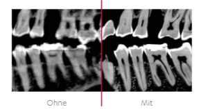
3Shape’s motion compensation technology minimizes blur and motion artifacts, delivering precise and clear images. 3Shape’s motion compensation is based on head tracking technology that measures patient movement while scanning. A powerful reconstruction software compensates all movements of the patient. The CBCT, panoramic slice and 3D facial scans are delivered clearly and precisely – motion artifacts are common in children or the elderly. This can lead to a reduced image quality and limit the diagnostic and treatment options. Furthermore, the motion compensation ensures that precise recordings can be made without artifacts.
Recording with and without motion compensation
Dynamic field of view
The dynamic field of view allows you to scan the most relevant areas in detail with a low radiation dose for patients.
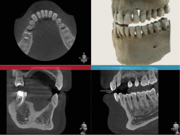
The combination of high-quality CBCT recordings, panoramic slice recordings and 3D facial scans using 3Shape X1 and 3Shape TRIOS intraoral scans allow precise diagnosis and treatment planning.

With 3Shape Digital Patient (intelligent patient management) patient data can be stored digitally in one place. This in turn enables a better follow-up examination.
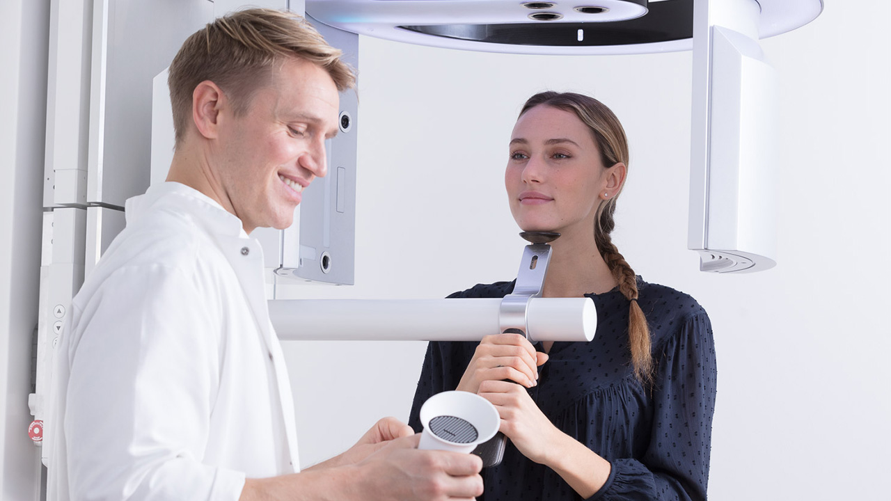
Benefit from integrated digital workflows with 3Shape X1 scans and 3Shape apps.
Are you already using 3Shape Implant Studio?
This tool allows you to offer digital implant planning to improve predictability and patient comfort.
You can improve the treatment results through prosthetic implant planning based on 3Shape X1 images and TRIOS intraoral scans. In addition, it enables better planning and uniform workflows for implant care thanks to digital accuracy. You also have the opportunity to benefit from guided surgery – either by designing and printing your own surgical guides on site or by sending your designs to an external partner.
