Product Highlights
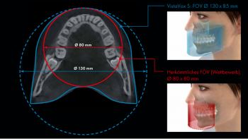
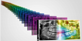
Jaw-shaped Field of View
The VistaVox S has a jaw-shaped volume that images the diagnostically relevant area of a Ø 130 x 85 mm volume. This volume is considerably larger than the conventional volume of Ø 80 x 80 mm and can thus also completely cover the region of the rear molars. By means of a special curved path with 540° rotation, the ideal jaw-shaped volume can be achieved. The VistaVox S needs only 18 seconds for the whole process. Together with a tightly collimated conical beam and the highly sensitive CsI sensor, this X-ray unit enables a particularly low radiation dose.
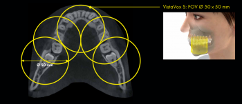
Ten additional volumes
In addition to the jaw-shaped volume, the VistaVox S has 10 further volumes with Ø 50 x 50 mm: five for the upper jaw and five for the lower jaw. These are used when only a very specific area of the jaw needs to be covered e.g. for endodontic and implant treatments.
Standard Quality Mode (SQ Mode)
The SQ mode makes it possible to reduce the X-ray dose even further as 62% less dose is used in this setting than in HQ mode (Highest Quality mode). This mode can be selected for all programmes. It is particularly suitable for implant planning, determining the apical bone status, for examining the sinuses or for locating impacted or excess teeth.
Panoramic X-ray programmes
The VistaVox S offers 17 X-ray programmes with which you are equipped for every diagnostic requirement. In addition to the standard panoramic programme, there are various programmes for children, programmes for orthogonal X-ray images, for temporomandibular imaging and for sinus X-ray images.
S-Pan technology
With this technology the image areas that best correspond to the patient anatomy are selected from a number of parallel layers. When these are combined, the result is a panoramic image. The reconstruction is based on the effective position of the bite, resulting in a clear image that also includes individually-angled teeth. This means that incorrect positioning can also be balanced out to a certain extent, which saves an enormous amount of time for the practice as well as for the patient.
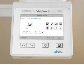
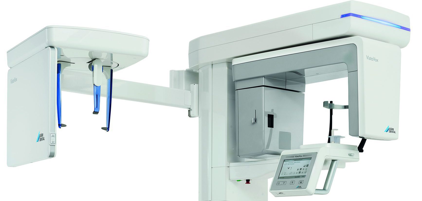
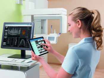
7″ touch display
With this innovative display, operating and navigating the VistaVox S has never been easier! This helps with a smooth workflow during the X-ray.
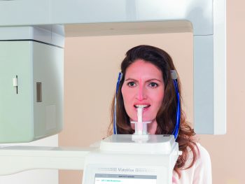
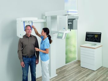
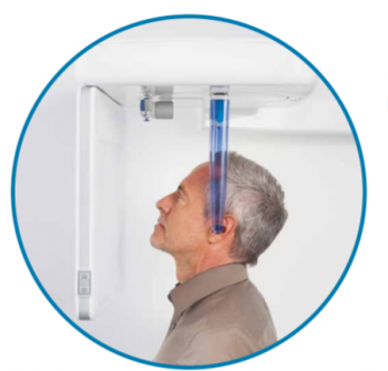
Patient positioning made easy!
With the help of three light lines (sagittal, Frankfurt plane and Canine) for 2D images and two light lines (sagittal and horizontal plane) for 3D X-ray images, it is very easy to position the patient correctly.